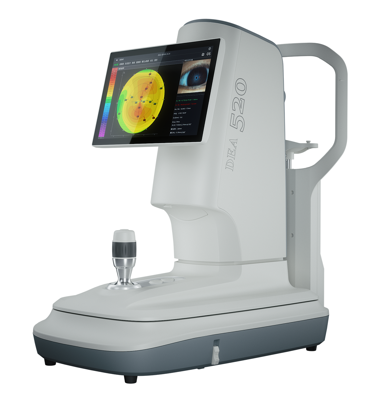
2 in 1 Ocular Diagnostic Master
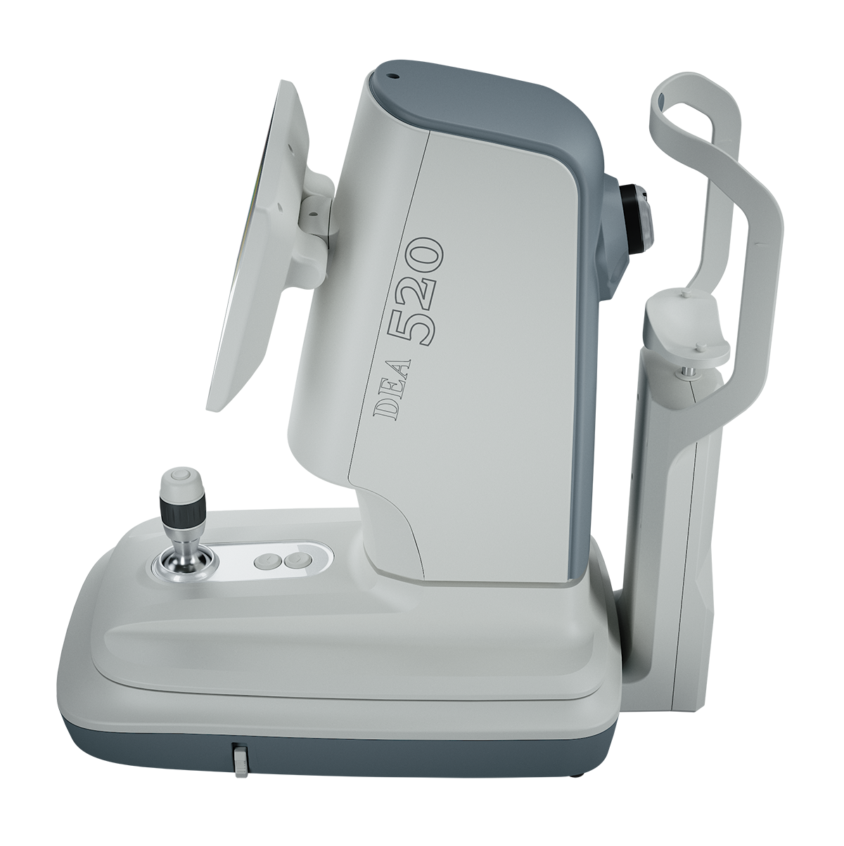
The Pathfinder is an advanced multi-purpose corneal topographer designed to streamline dry eye diagnostics and corneal topography analysis. Featuring a built-in computer, it maximizes exam room efficiency while integrating essential diagnostic capabilities. Its user-friendly design enhances doctor-patient communication with easy-to-understand visual reports and real-time observation via external display connection. The ergonomic 50° adjustable display, intelligent auto eye recognition, and compact cone ensure precise operation and adaptability. With versatile clinical applications, including dry eye analysis, corneal morphology diagnosis, aberration simulation, and lens fitting, the Pathfinder is an indispensable tool for modern ophthalmic care.
Pathfinder Corneal Topographer Brochure
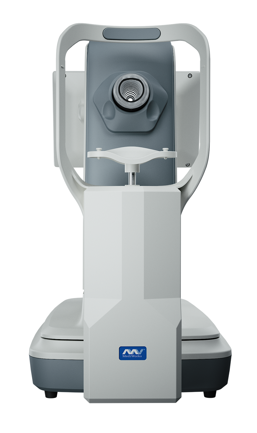
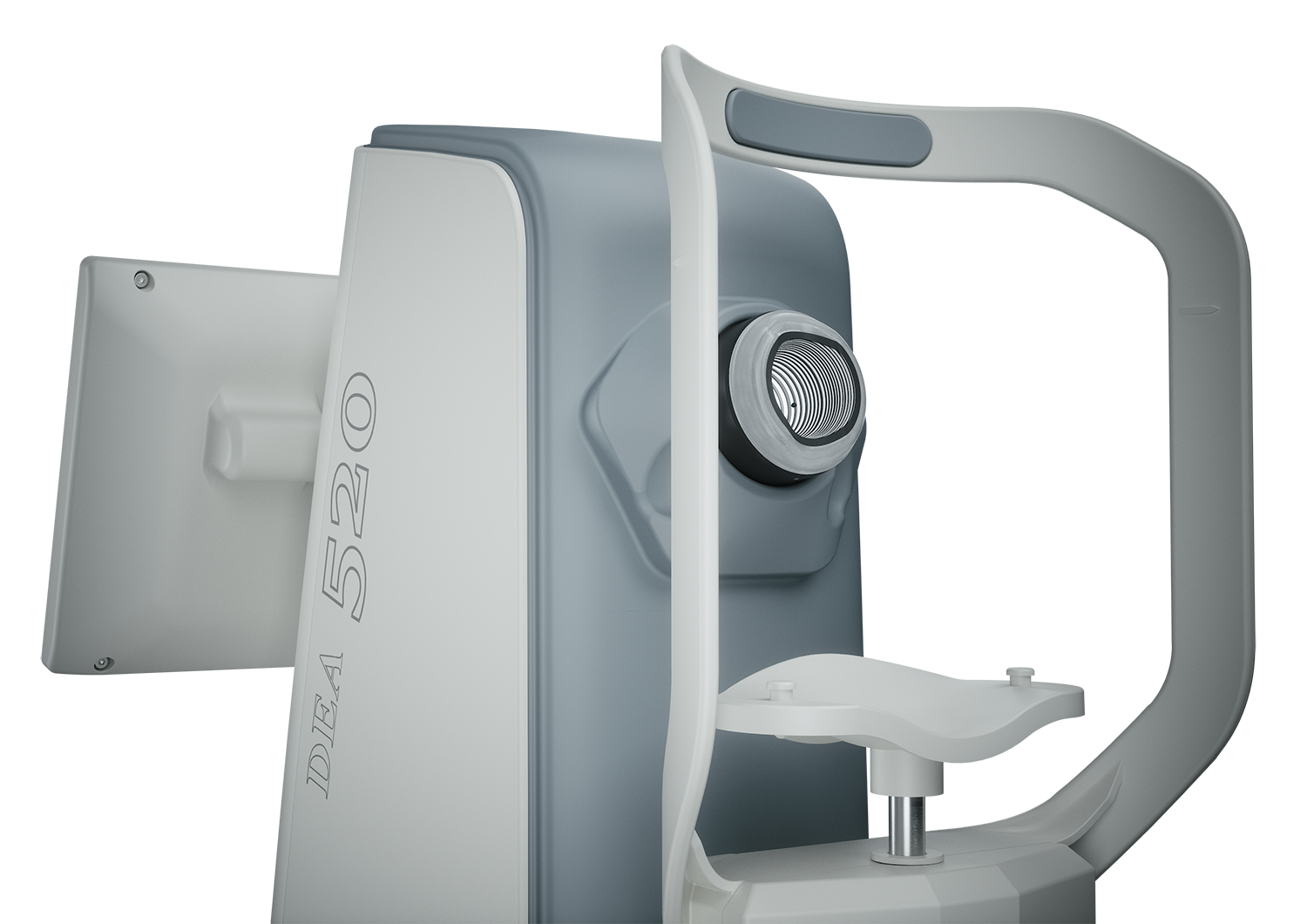
Placido Ring
- Thousands of measuring points – ensures more data is available with accurate analysis
- Smaller cone design – bigger projection area
- 3 Illuminations – white illumination, infrared illumination, cobalt blue illumination
14 Functions
Dry Eye Diagnostics
- Dry Eye Questionnaire
- Non-Invasive Tear Film Breakup Time
- Conjunctival Redness Analysis
- Meibomian Glands Function Evaluation
- Cornea Sodium Fluorescein Staining
- Non-Invasive Tear Meniscus Height
- Lipid Layer Thickness
- Blink Quality
- Eyelid Margin
- Fluorescein Tear Film Breakup Time
Topography
- Topography Analysis
- Lens Fitting
- Aberration & Simulation
- Pupil & Corneal Diameter Measurement
Dry Eye Diagnostics
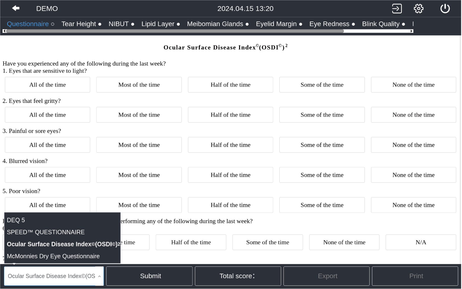
Dry Eye Questionnaire
Ocular Surface Disease Index (OSDI)/McMonnies/SPEED/DEQ 5
The built-in dry eye questionnaire is designed according to the risk factors and clinical characteristics of dry eye. It offers a simple preliminary dry eye assessment, enhancing diagnostic accuracy, treatment efficiency, and patient follow-up care.
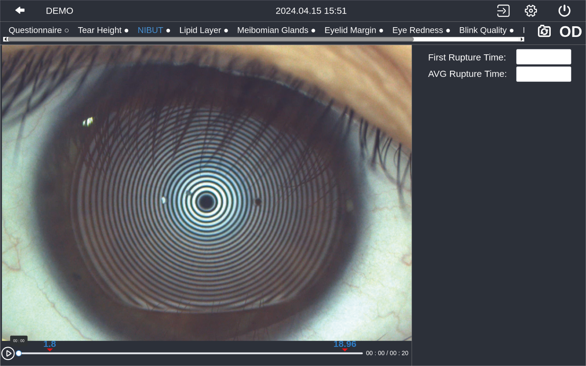
Non-Invasive Breakup Time
Interface - Comprehensive dry eye examinations.
NIBUT - More than 9.6 mm diameter Placido ring projection. Auto-identify the breakup area and analyze NIBUT intelligently.
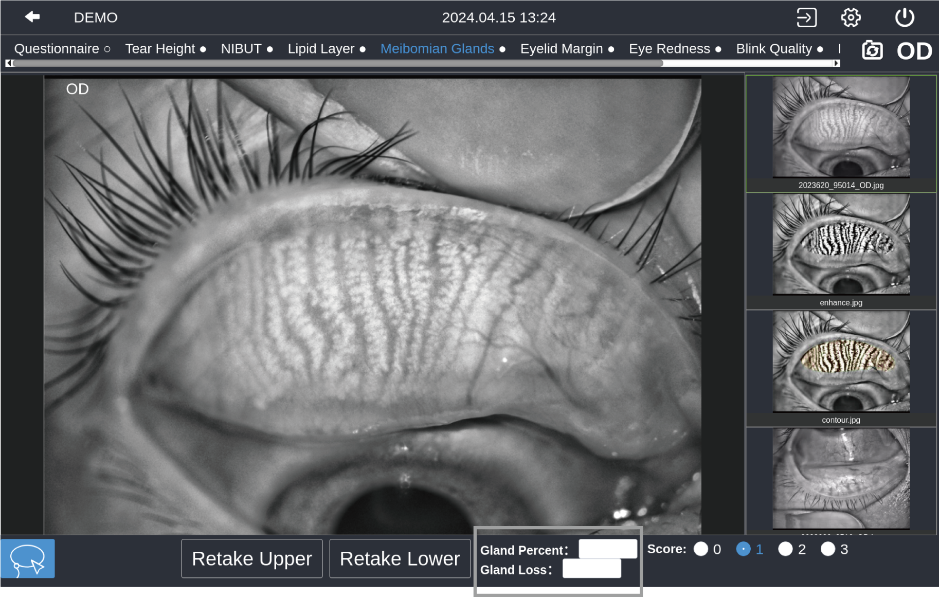
Meibomian Glands Function Evaluation
Auto-identify and auto-enhance meibomian gland images
Automatically analyzes meibomian glands loss caused by dysfunction of the meibomian glands, providing accurate and quantified diagnostic results.
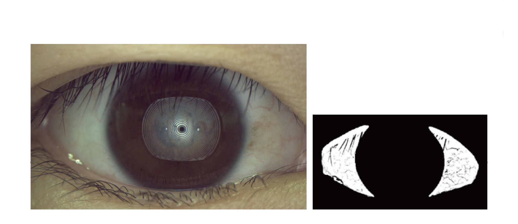
Non-Invasive Tear Meniscus Height
Automatic identification system depicts tear meniscus area and measures the tear height intelligently.
Conjunctival Redness Analysis
Identify and calculate percentages of conjunctival congestion, ciliary congestions, and evaluate severity of eye congestion.
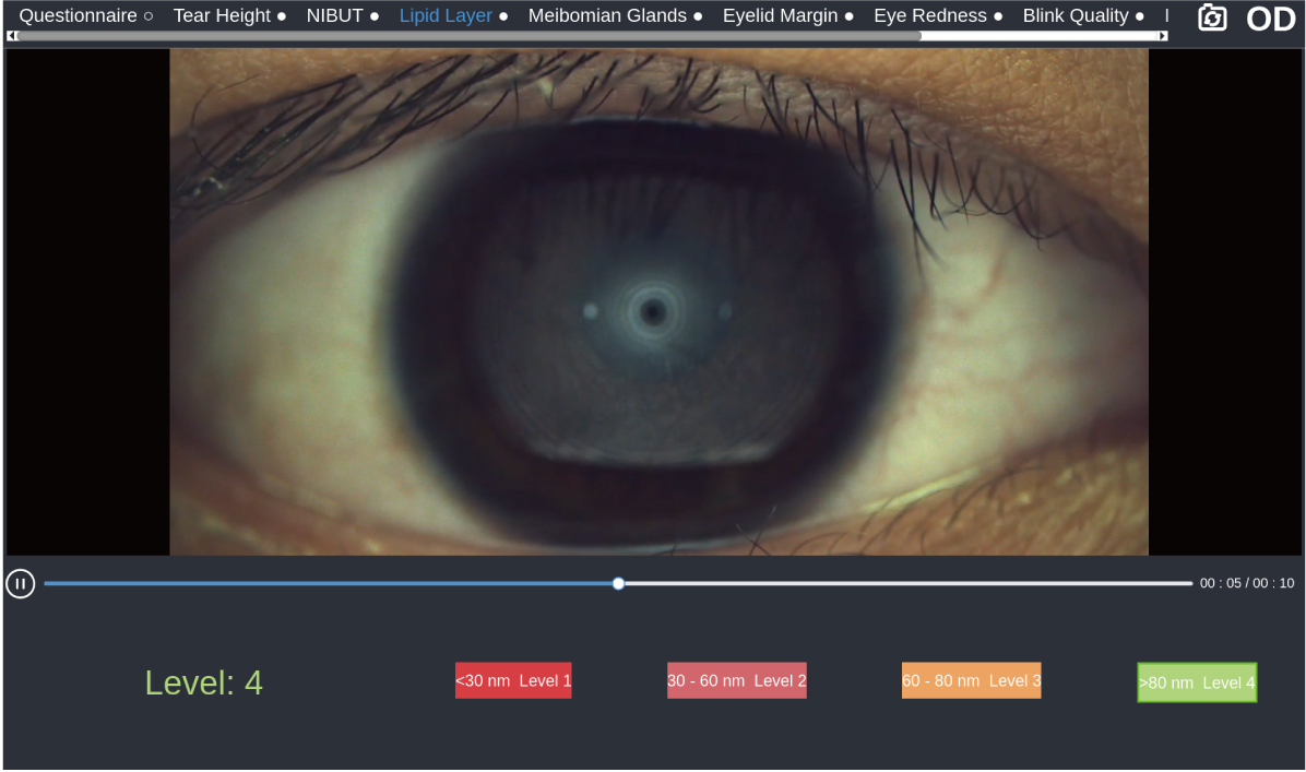
Lipid Layer Thickness
Monitor the dynamic lipid layer and its distribution through video recordings compared to standard templates, aiding in the assessment of MGD.
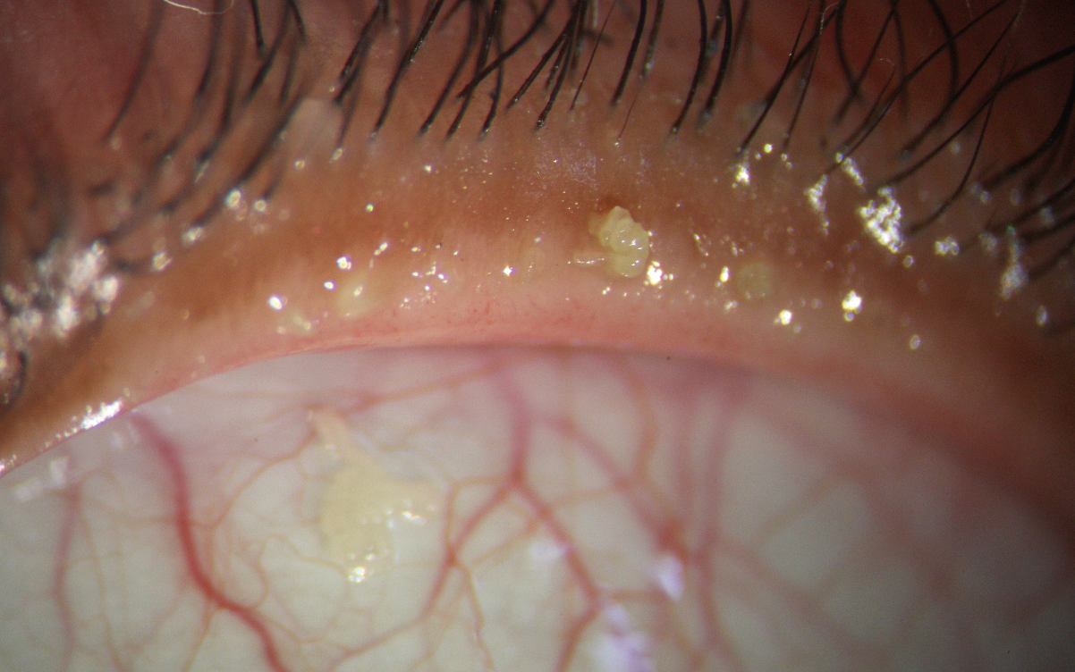
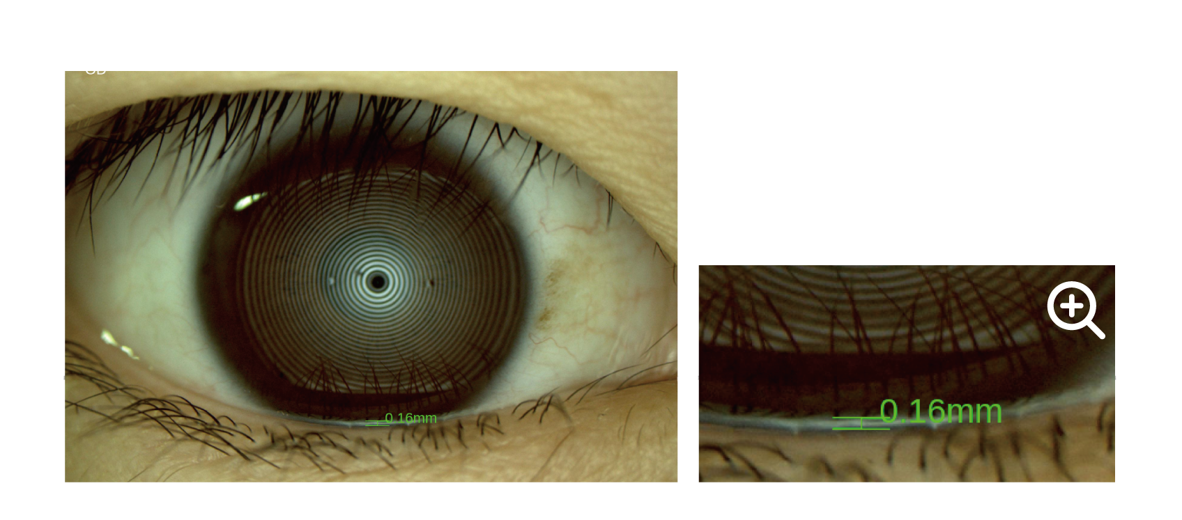
Eyelid Margin
The high-resolution image allows for zooming, enabling detailed examination of the overall shape of the eyelid margin and subtle changes.
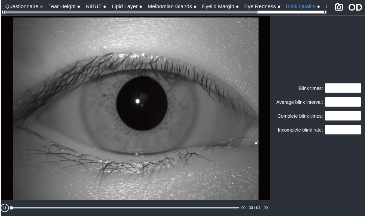
Blink Quality
High-definition video is captured with an infrared light source and automatically analyzes blink frequency, blink interval, incomplete blink, and incomplete blink ratio.
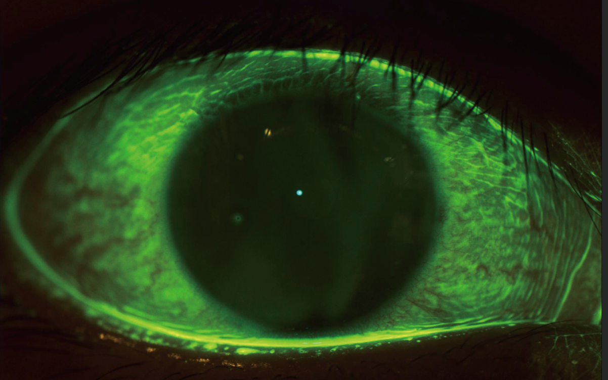
Corneal Fluorescein Staining
The specially designed built-in yellow filter, combined with cobalt-blue illumination, enhances the contrast of corneal fluorescein staining images, effectively increasing the detection rate of early corneal epithelial staining.
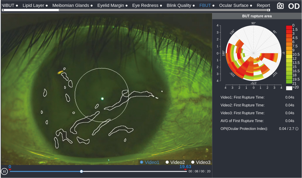
Fluorescein Tear Film Breakup Time
AI technology automatically detects changes in tear film morphology and calculates tear film breakup time to assess tear film stability.
Corneal Topography
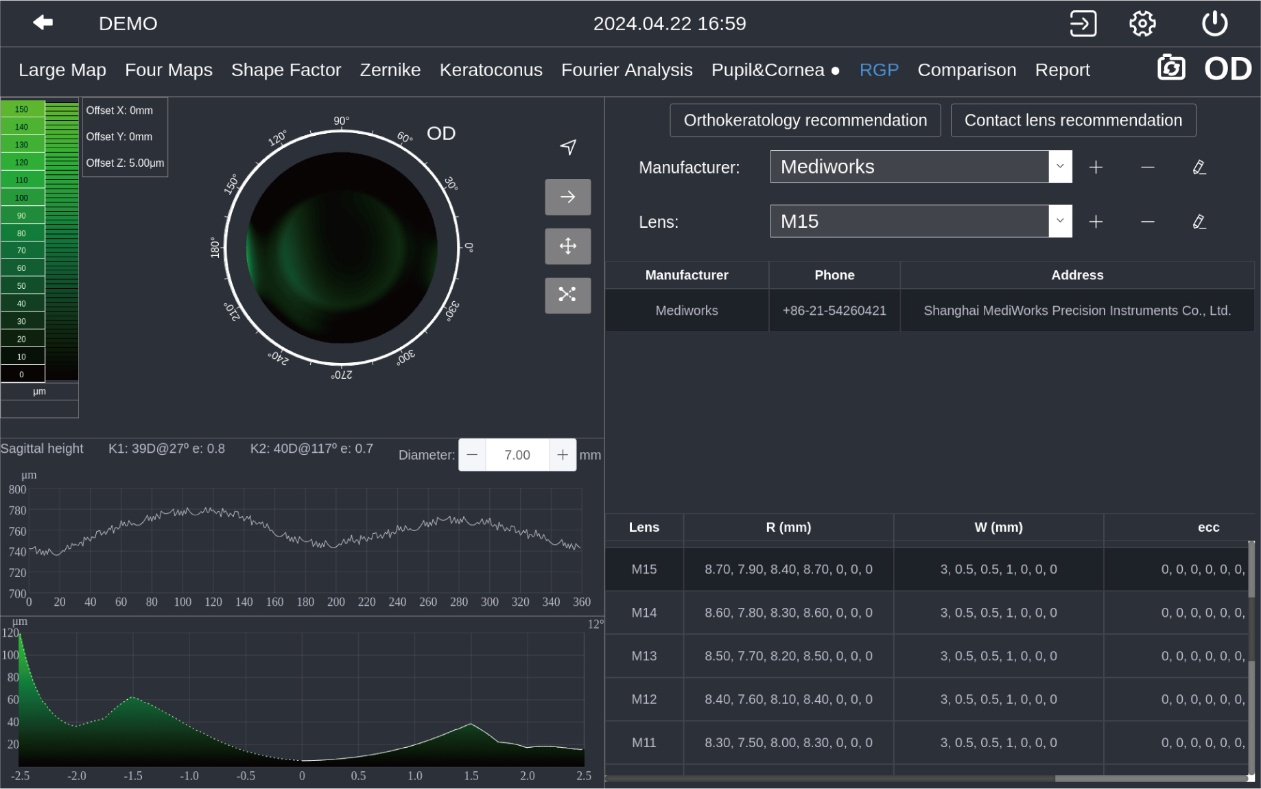
Lens Fitting
A simulated fluorescein image will be created based on patient's cornea. The system will recommend several suitable lenses to choose, which accelerates the workflow and excludes unfit lens to save the trouble for patient to do real several fluorescein staining.
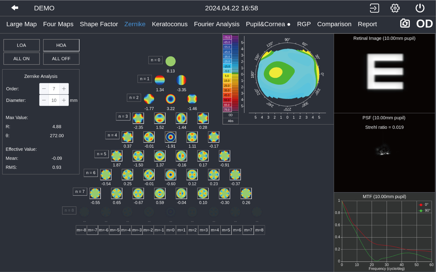
Aberration & Simulation
Zernike wavefront aberration analysis visualizes cataract and refractive surgery planning, ensuring optimal postoperative vision quality for patients.
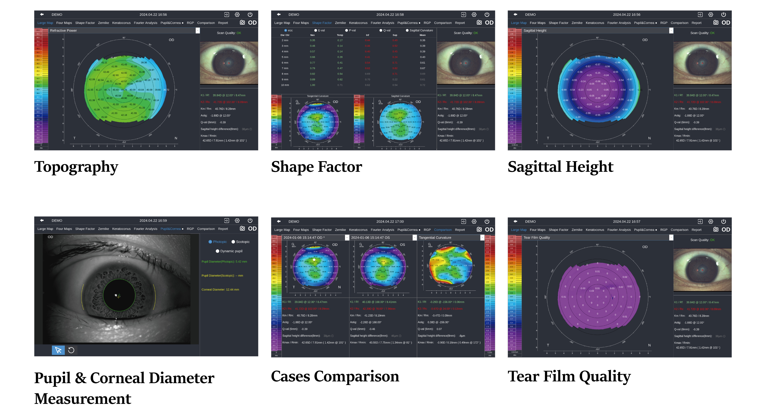
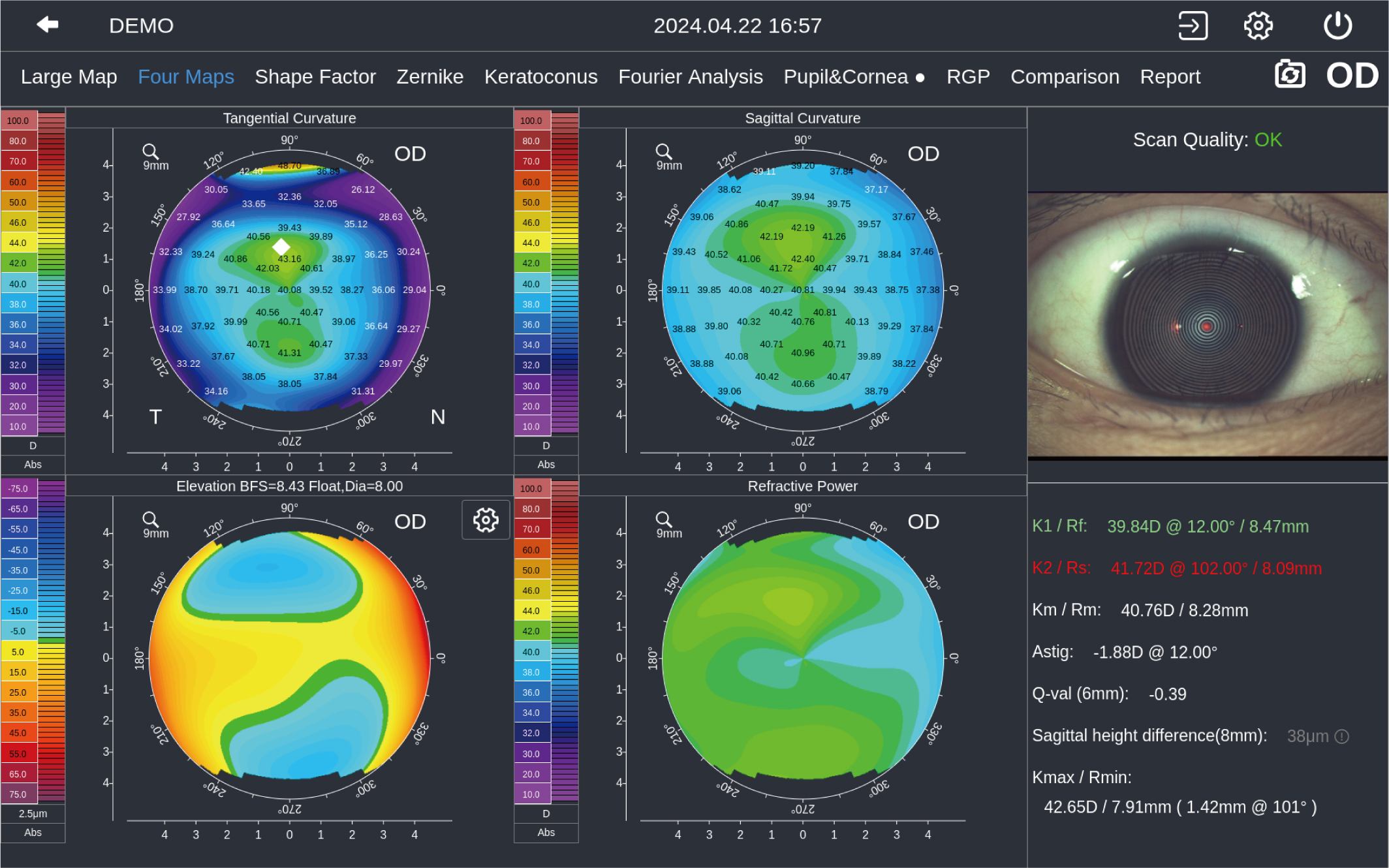
Maps
Maps provide Sagittal Curvature, Tangential Curvature, Elevation Map, Refractive Power, K1/K2/Km/Astig/Ecc value, and more!
Send me more info on the Pathfinder Corneal Topographer
Specifications
| Hardware | |
|---|---|
| Dimension | 49 cm × 31 cm × 54 cm |
| Weight | 16.1 kg |
| Built-in CPU | intel |
| Hard Disk | 1 TB |
| Image Resolution | 2048 × 1536 |
| Display | 10.1″ touchscreen |
| Illumination | White, Infrared, Cobalt-blue |
| Internet Connection | Wifi & Wired network |
| Printer Connection | WIFI, USB |
| Power Supply | 100 ~ 240 VAC, 50 / 60 HZ |
| Extension Display Interface | Display Port |
| OS/OD Recognition | Automatic |
| Chin Rest Control | Electrical |
| Left and Right | 0 ~ 90mm work range |
| Front and Back | 0 ~ 60 mm work range |
| Up and Down | 0 ~ 30 mm work range |
| Language | Chinese / English / Japanese / German / Italian |
| DICOM | Supported |
| Topography | |
| Numbers of Rings | 50 Rings |
| Diameter of Project | Area 8.8 mm (42.18 D) |
| 11 mm (42.18 D) | |
| Radius of Curvature | 32.14 dpt ~ 61.36 dpt (5.5 mm ~ 10.5 mm) |
| Accuracy: ± 0.1 dpt (± 0.02 mm) | |
| Astigmatism Axis | 0 ~ 180° |
| White To White | 1 ~ 20 mm |
| Pupil Diameter | 1 ~ 13 mm |
| Topography Function | Sagittal Curvature |
| Tangential Curvature | |
| Elevation Map | |
| Refractive Power | |
| Sagittal Height | |
| Tear Film Quality | |
| 4 Maps | Four Maps display |
| Shape Factor | Ecc, E, p, Q |
| Zernike | Corneal wavefront aberration, PSF map, MTF curve and Simulated image in different pupil diameters |
| Examination Result Comparison | Support 2 results comparison and difference calculation |
| Dry Eye Analysis | |
| NIBUT | Automatic analysis, tear film rupture area and trend, first break-up time and average break-up time |
| Tear Meniscus Height | 0.01 ~ 2 mm |
| Meibomian Glands | Meibomian glands loss rate and grade |
| Lipid Layer | Template match |
| Eye Redness | Conjunctival congestion percentage |
| Eyelid Margin | Support digital images zoom in |
| Ocular Surface | Built-in yellow filter |
| Blink Quality | |
| Fluorescein Tear Film Breakup Time | |